This piece of high-tech equipment is used in Biology and Metallurgy courses
It sits quietly in Room 175 of the Biomedical Technology building, exuding a formidable presence even when not in operation. Weighing more than 1,800 pounds – nearly a ton – it stands almost 6 feet tall on its pedestal and is approximately 2 ½ feet long and 3 feet deep.
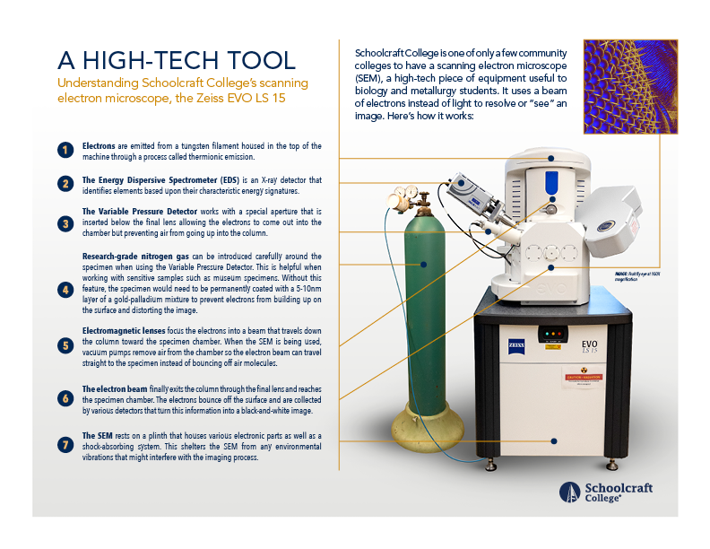
In the scientific and technical community, it’s called the Zeiss Evo LS 15. Less formally, it’s the College’s scanning electron microscope and has been here since 2008.
Melissa Gury, Director of Laboratory Sciences, has been working with the scanning electron microscope, or SEM, since 2014. She’s eager to share its abilities with an even wider audience.
“It’s very rare for a community college to have such a high-tech, industry-standard piece of equipment like this,” Gury said. Gury said she didn’t know of any area community colleges that have an SEM and that some four-year schools and even businesses seek out Schoolcraft to use it.
What is an SEM?
Unlike the microscope that you might have used in high school biology, a scanning electron microscope uses a beam of electrons instead of light to “see” an image. The process, as explained by Gury, works like this:
- SEMs contain electromagnetic lenses that focus the beam of electrons, which is passed across the surface of a specimen.
- Electrons bounce off the surface of the specimen and are collected by various detectors that turn this information into a black-and-white image.
- Vacuum pumps physically remove air from the chamber so that the electron beam can travel straight to the specimen instead of bouncing off air molecules.
- This normally means that to use a traditional detector, the specimen has to be completely dried.
- If it is not conductive, it has to be coated in a very small (5-10 nm) layer of a gold-palladium mixture.
- Schoolcraft’s SEM has a Variable Pressure Detector and a special aperture that is inserted below the final lens allowing the electrons to come out into the chamber but preventing air from going up into the column.
- This allows the SEM to introduce a small amount of research-grade nitrogen gas around the surface of the specimen, meaning the specimen does not have to be completely dry or covered with any other materials.
- This can be especially helpful when working with something like a museum specimen or some other item that is rare so it is not harmed during the imaging process.
“Typically, at a university, a student would have to be in a 400-level course or even in graduate school before using a scanning electron microscope.”
Melissa Gury, Director of Laboratory Sciences
What classes use it?
Two classes currently use the SEM: BIOL 140 (Scanning Electron Microscopy) and MET 248 (Metallurgy and Materials Science).
Here’s the course description for BIOL 140, which does not require a prerequisite:
This course emphasizes the principles and modes of operation of the scanning electron microscope and X-ray analysis systems, electron-specimen interactions, elemental analysis, effects of microscope variables on images, image processing, routine maintenance, the use of microscope accessories and digital outputs. In the laboratory, students will prepare and examine inorganic and organic specimens using the secondary, backscatter and variable pressure detectors of the SEM. Students complete a project consisting of the preparation, imaging and analysis of a biological specimen.
Photo 1 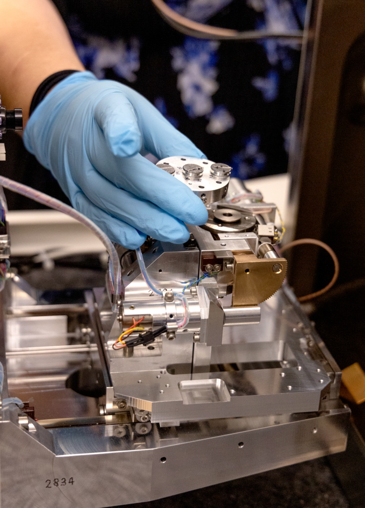
Photo 2 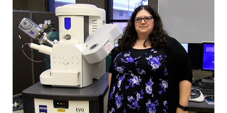
Photo 3 
Photo 4
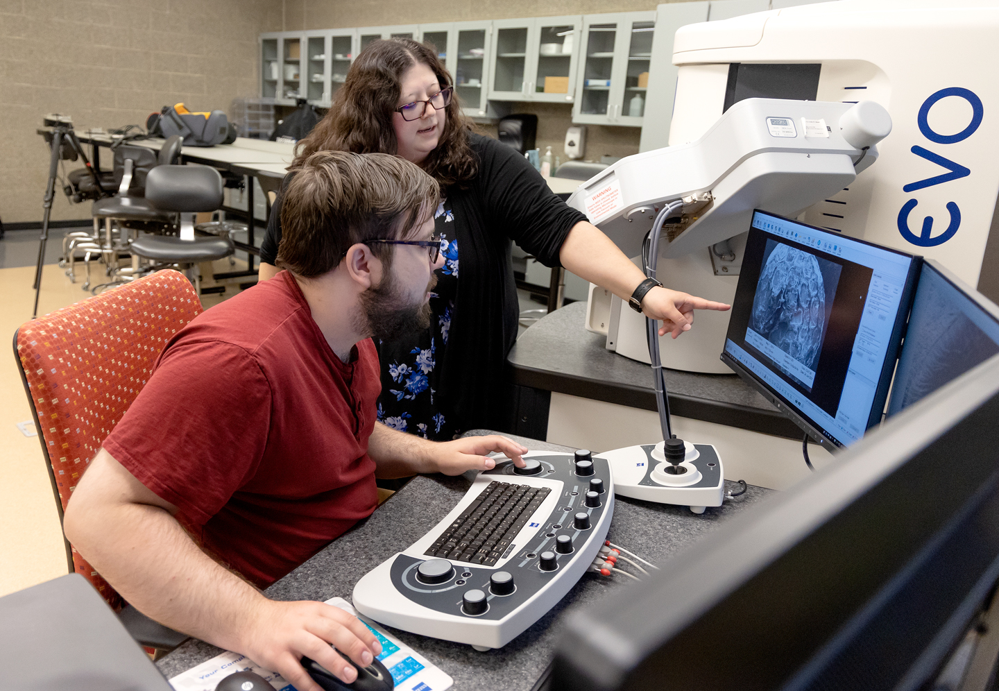
Photo 2: A specimen is placed in the scanning electron microscope in the Biomedical Technology building at Schoolcraft College.
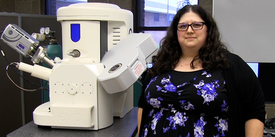
Photo 3: Melissa Gury, Director of Laboratory Sciences, with Schoolcraft College’s scanning electron microscope.
Photo 4: Melissa Gury instructing: Melissa Gury, Director of Laboratory Sciences, instructs on how to examine a specimen in the scanning electron microscope at Schoolcraft College.
“Typically, at a university, a student would have to be in a 400-level course or even in graduate school before using a scanning electron microscope,” Gury said. “With BIOL 140, we’re able to expose our students to amazing technology early in their academic path.”
Here’s the course description for MET 248, which requires MET 153 or department consent:
This course introduces the fundamentals of Scanning Electron Microscopy (SEM) and X-ray Microanalysis used for materials characterization and failure analysis. Topics include microscopy systems and components, safety and maintenance, applications in fractography and materials characterization and failure analysis.
Who else could use it?
Biology researchers as well as businesses and industries connected to metallurgy, geology, forensics and more all could find Schoolcraft College’s scanning electron microscope highly useful. For example, Schoolcraft College is currently assisting an area university student with research into diseases that affected amphibians. (The disease does not affect humans.) The student is able to examine rare museum specimens in a nondestructive manner to perform the research.
Business and industry that need to analyze parts or assemblies for fractures rely on scanning electron microscopes as well. The SEM can analyze the elemental composition, though not the actual “recipe” of components.
“Our scanning electron microscope is another example of how Schoolcraft College is providing the equipment and training to prepare our students for high-tech careers,” Gury said.